Real-Time Metabolic Imaging. Made Simple.
On-site, push-button production of metabolic imaging agents
Real-Time Metabolic Imaging in 3 Simple Steps

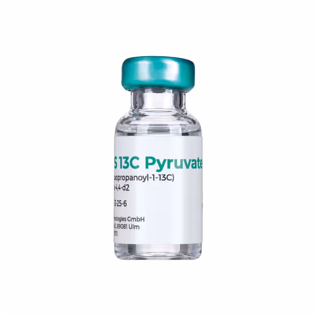
Prepare
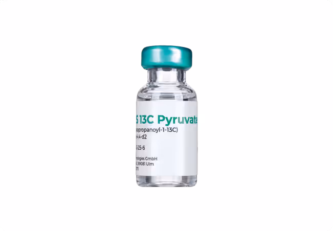
Take a precursor metabolic agent (organic molecule natural to the body), e.g., 13C enriched pyruvate

Polarize
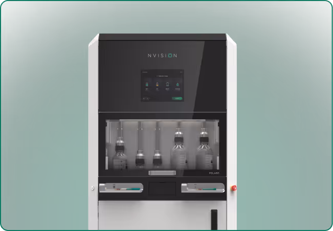
Use POLARIS to amplify the MRI signal of the metabolic agent by >10,000x through spin hyperpolarization

Scan
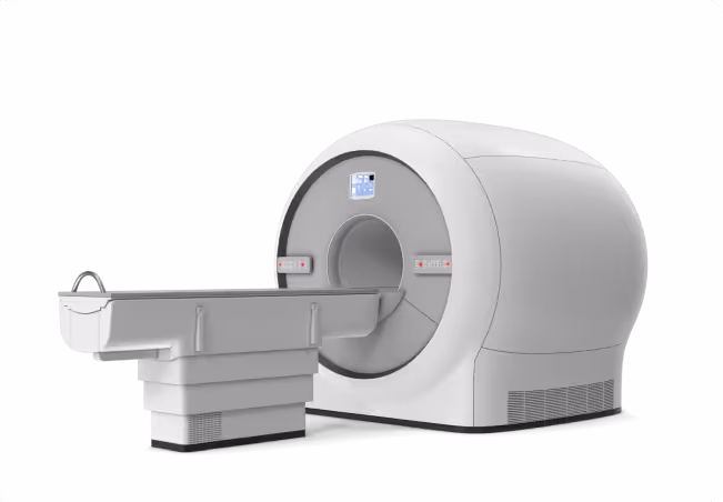
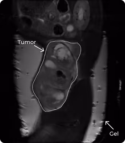
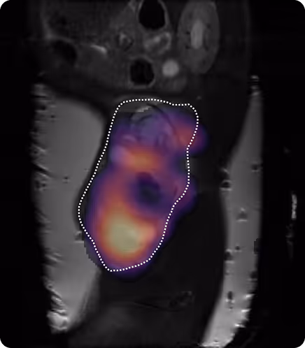
Inject the hyperpolarized agent and measure its metabolism using standard MRI

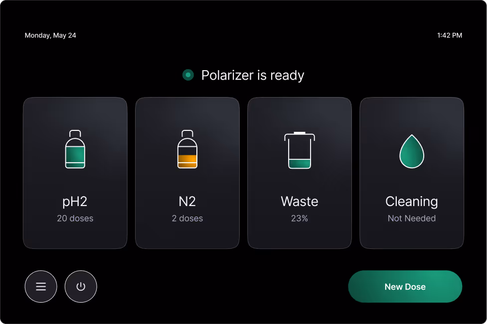
POLARIS minimizes variability through standardized kits and automated polarization and purification.
Explore how POLARIS could fit into your workflow
Key Benefits
- High throughput (up to 3 doses/hour)
- Convenient, room temperature processing
- Small footprint, minimal infrastructure adjustment
- Simple, robust workflow with ready-to-use kits
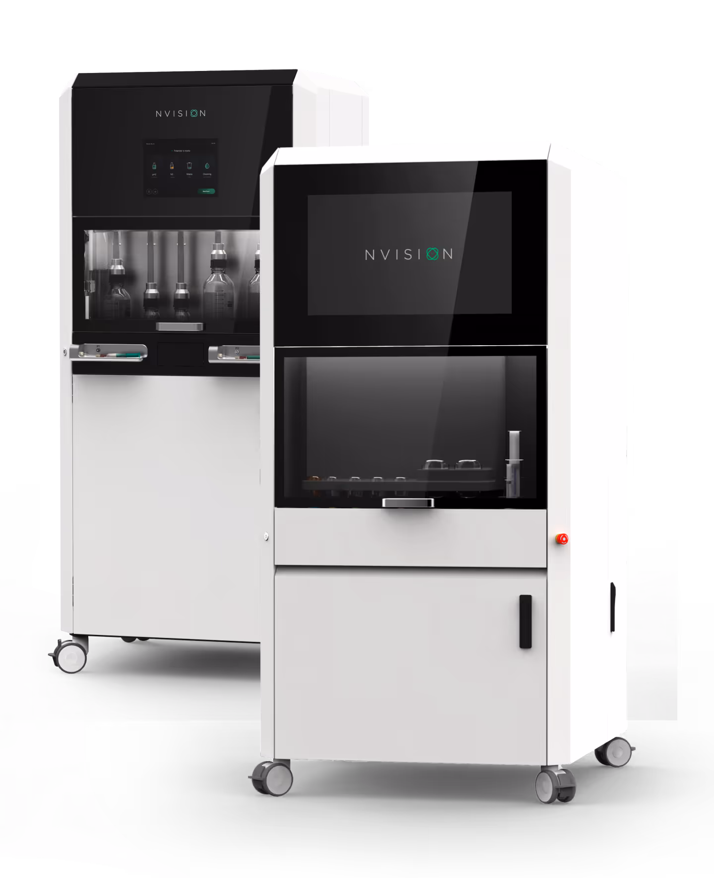
An End-to-End Solution for Rapid Setup in Your Lab
Polarizer
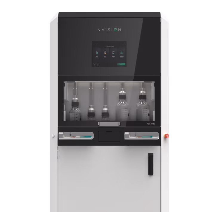
Consumables

Support
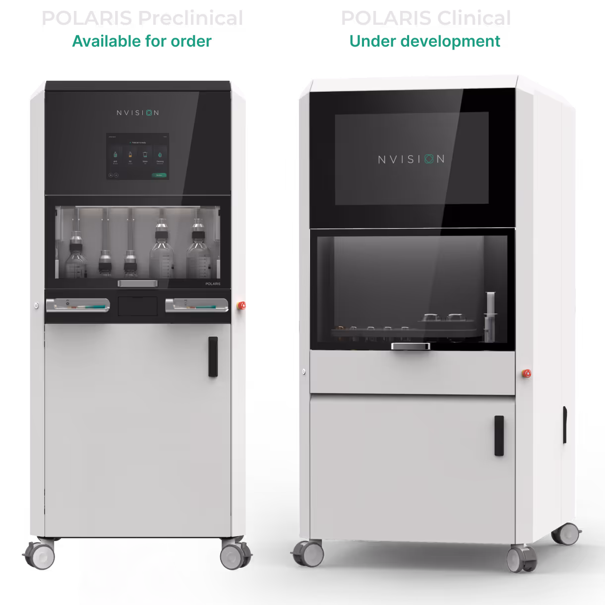
Experience with POLARIS in Preclinical Use
"POLARIS joined our lab like a shooting star! Being able to quickly obtain a strong and reproducible hyperpolarized pyruvate signal was our dream for a long time.
POLARIS Preclinical is integrated in our standard preclinical metabolic imaging workflows with hyperpolarized pyruvate in rodent studies. The system's ease of use and small footprint support ongoing preclinical studies in oncology, physiology and neurology.
We are looking forward to continue working with it together with the incredible team from NVision!"
Technical University of Munich
Obtain High-Quality Hyperpolarized Signal With PHIP
POLARIS uses parahydrogen-induced polarization (PHIP) to produce hyperpolarized metabolic imaging agents. PHIP enables strong, robust signal enhancement for real-time metabolic imaging within a streamlined, high-throughput workflow.