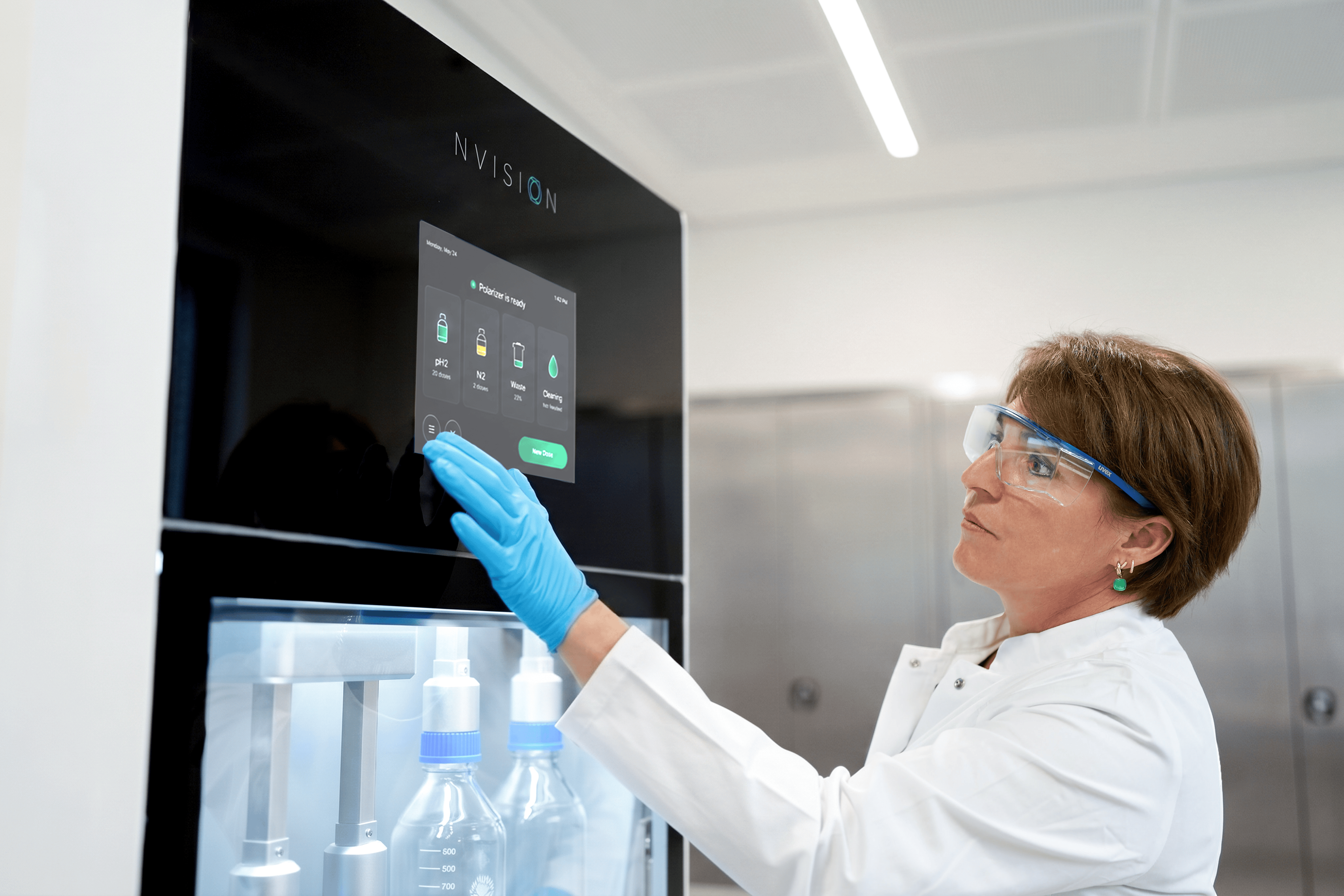

Scientist operating the POLARIS hyperpolarization system in the laboratory

Gain Critical Metabolic Insights Across Diseases and Organs

  • In vivo
    Validation
  • Metabolic
    Phenotyping
  • Treatment
    Response Biomarker
  • Enable Clinical
    Translation

Explore disciplines

Oncology

Characterize tumor models and predict responses to novel interventions in vivo

Metabolic imaging distinguishes ovarian cancer subtypes and detects their early and variable responses to treatment. Chia et al. 2024. Oncogene.

Challenge

Leverage HP MRI data to empower your basic biomedical research

Approach

Map these critical metabolic fluxes in vivo with metabolic imaging agents

Outcome

Use these real-time insights to precisely characterize tumor models and predict responses to novel interventions

Drug Development

Adding a dynamic metabolic readout to existing disease models significantly accelerates the optimization of new therapies. Our robust hyperpolarized MRI technology complements traditional assays by delivering actionable in vivo validation.

Accelerate your pipeline with rapid, translatable insights:

Establish Baselines

Non-invasively map the metabolic profile of your models prior to drug administration.

Confirm Pharmacodynamics & Target Engagement

Obtain in vivo mechanistic information, directly observing target pathways to determine if treatment normalizes metabolic phenotypes before clinical outcomes emerge.

Optimize Protocols

Proactively refine dosing, schedules, and drug combinations based on immediate metabolic data.

Identify Optimal Timing of Interventions

Detect early-stage disease to narrow down the most effective window for treatment initiation to maximize therapeutic impact.

Monitor in Real-Time

Track treatment responses and gain actionable feedback on drug efficacy within days.

De-Risk Development

Utilize the exact same translatable readouts from early in vitro experiments through preclinical models and into clinical trials.

NVision’s quantum technologies provide an end-to-end platform for drug development:

Quantum computing chip for simulating molecular interactions

Quantum Computing (PIQC)

could simulate drug-target interactions with greater accuracy, leading to the discovery and design of better candidates

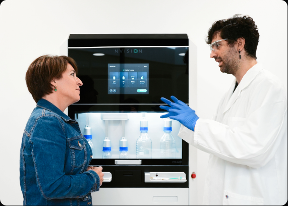
Two researchers discussing the POLARIS system in a laboratory

Quantum-Enhanced MRI (POLARIS)

could rapidly validate a candidate’s efficacy in a native biological environment

By bridging the gap between in silico design and in vivo testing, these two synergistic technologies will accelerate drug development and address previously undruggable targets.

In Vitro

Measure pharmacodynamic effects in vivo

In Vivo

Access efficacy earlier

Clinical

Optimize treatment timing with early disease biomarkers

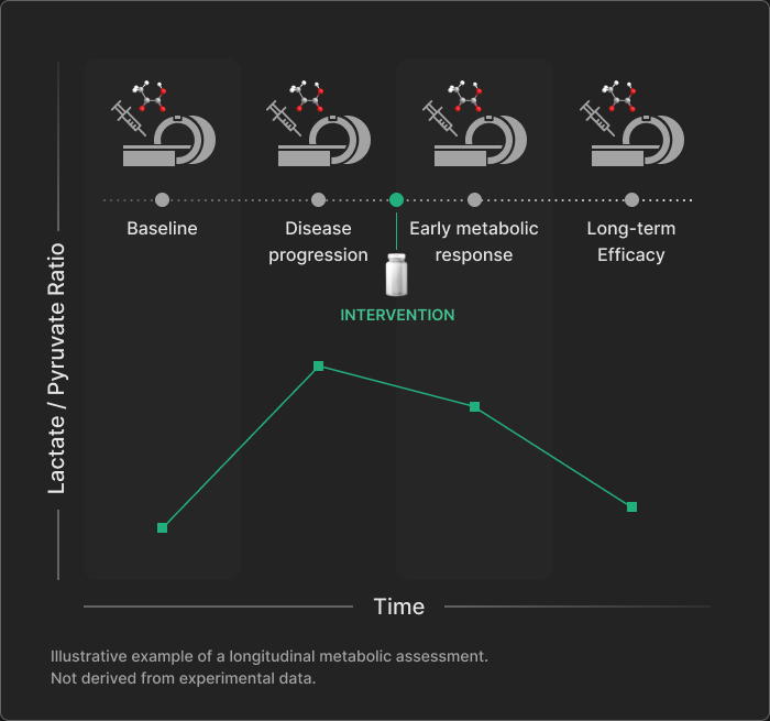
Longitudinal Metabolic Imaging

Illustrative longitudinal metabolic assessment: the lactate-to-pyruvate ratio rises from baseline during disease progression, then declines following intervention through early response and long-term efficacy. Not derived from experimental data.

Quantify real-time metabolic flux in the same subject from baseline through disease progression and therapeutic intervention.

Overcome the reliance on terminal endpoints by leveraging repeated measurements across the disease continuum. This enables intra-subject comparisons at every stage, enhancing the robustness and translational value of preclinical studies.

Cardiology

Uncover cardiac energetic shifts and monitor treatment responses across heart failure, ischemia, and metabolic syndrome in vivo

Abnormal energy metabolism drives obesity-related heart dysfunction and cardiovascular risk, yet accurately assessing these metabolic perturbations non-invasively remains a challenge.

Metabolic MRI was used to track the conversion of hyperpolarized [1-13C]pyruvate to assess real-time changes in fundamental cardiac carbohydrate use.

Metabolic MRI provided a non-invasive way to detect obesity-driven cardiac dysfunction and track the restorative effects of dietary and pharmacological treatments.

Hyperpolarized 13C and 31P MRS detects differences in cardiac energetics, metabolism, and function in obesity, and responses following treatment
Lewis et al.
2024
NMR Biomed.
Early detection of doxorubicin-induced cardiotoxicity in rats by its cardiac metabolic signature assessed with hyperpolarized MRI
Timm et al.
2020
Commun Biol.

Hyperpolarized [1-13C]pyruvate detects early drug-related toxicity in the heart before functional deficits manifest.

Hyperpolarized [1-13C]pyruvate cardiovascular magnetic resonance imaging identifies metabolic phenotypes in patients with heart failure
Joergensen et al.
2024
J Cardiovasc Magn Reson.

By identifying a distinct metabolic signature at the time of revascularization, metabolic imaging agents enable the prediction of functional recovery one year later in patients with cardiac ischemia.

Neurology

Monitor disease onset and validate novel therapeutic strategies for neurodegenerative and neuroinflammatory conditions

Secondary injury after traumatic brain injury due to mitochondrial damage, hypoxia, and inflammation resulting in metabolic shifts are difficult to identify, limiting the ability for early detection and timely intervention.

Metabolic MRI was used to track the conversion of hyperpolarized [1-13C]pyruvate to both bicarbonate and lactate to assess real-time shifts in cerebral metabolism.

Metabolic MRI identified aberrant mitochondrial metabolism as an in vivo biomarker to track secondary injury progression in traumatic brain injury models.

Longitudinal assessment of mitochondrial dysfunction in acute traumatic brain injury using hyperpolarized [1-13C]pyruvate
Hackett et al.
2023
Magn Reson Med.
Imaging immunomodulatory treatment responses in a multiple sclerosis mouse model using hyperpolarized 13C metabolic MRI
Guglielmetti et al.
2023
Commun Med (Lond).

Hyperpolarized [1-13C]pyruvate shows potential for non-invasive monitoring of immunomodulatory treatments in a multiple sclerosis model, detecting a decrease in neuroinflammation not detectable with conventional imaging.

Age-associated change in pyruvate metabolism investigated with hyperpolarized 13 C-MRI of the human brain
Uthayakumar et al.
2023
Hum Brain Mapp.

By characterizing the metabolic landscape of the healthy aging brain, this study establishes a vital baseline to help future research distinguish normal age-related changes from the pathological signatures of neurodegeneration.

Redox Biology

Non-invasively assess oxidative stress and characterize cellular redox balance in vivo 

Invasive tissue extraction inherently disrupts the biological environment, providing limited ability to observe dynamic cellular redox states in their natural setting.

Metabolic MRI was used to track the NADH-dependent conversion of hyperpolarized [1-13C]pyruvate to lactate to non-invasively monitor the cellular redox state.

Metabolic MRI measured in vivo cellular redox states to reveal early diabetic kidney damage before changes appeared in routine clinical markers.

Multi-compartment metabolic assessment of the kidneys by co-hyperpolarized 13C MRI
von Morze et al.
2025
Magn Reson Med.
Assessment of Metformin-Induced Changes in Cardiac and Hepatic Redox State Using Hyperpolarized[1-13C]Pyruvate
Lewis et al.
2016
Diabetes.

By tracking shifts in the lactate-to-pyruvate ratio, this study demonstrates the ability of hyperpolarized [1-13C]pyruvate to non-invasively detect metformin-induced changes in the cytosolic redox state of the heart and liver in vivo.

Hepatology

Track early metabolic shifts associated with liver disease in vivo and evaluate the efficacy of novel interventions

Diagnosing metabolic liver disease currently relies on liver biopsies or structural imaging, offering limited access to the early, non-invasive biomarkers necessary for timely intervention.

Metabolic MRI was used to track the rapid conversion of hyperpolarized [1-13C]pyruvate, providing a non-invasive measure of hepatic metabolism over time.

Metabolic MRI identified alanine and lactate as non-invasive early biomarkers for hepatosteatosis in fatty liver disease.

Metabolic biomarkers for non-alcoholic fatty liver disease induced by high-fat diet: In vivo magnetic resonance spectroscopy of hyperpolarized [1-13C] pyruvate
Moon et al.
2017
Biochem Biophys Res Commun.
In vivo hyperpolarized carbon-13 magnetic resonance spectroscopy reveals increased pyruvate carboxylase flux in an insulin-resistant mouse model
Lee et al.
2013
Hepatology.

Hyperpolarized [1-13C]pyruvate detects increased gluconeogenesis in the diabetic liver and reveals a normalization of hepatic glucose metabolism after treatment with metformin.

In vivo Hyperpolarized Metabolic Imaging to Monitor the Progression of Hepatitis B Virus (HBV)-Related Hepatitis to Liver Fibrosis
Moon et al.
2024
Mol Imaging Biol.

Hyperpolarized [1-13C]pyruvate offers a non-invasive method to monitor the progression of HBV-related liver disease by detecting significantly elevated production of lactate and alanine, successfully distinguishing between normal liver tissue, hepatitis, and liver fibrosis.

Radiation Oncology

Non-invasively evaluate tumor radiosensitivity, monitor early treatment responses, and validate novel strategies for radiosensitization and sparing healthy tissue

Conventional imaging primarily tracks changes in tumor size, a delayed metric that might fail to capture early responses to radiation therapy.

Metabolic MRI was used to track the rapid conversion of hyperpolarized [1-13C]pyruvate to lactate, providing a real-time readout of altered cellular metabolic flux.

Metabolic MRI identified early, dose-dependent responses to radiation therapy in a prostate cancer model.

Simultaneous Metabolic and Perfusion Imaging Using Hyperpolarized 13C MRI Can Evaluate Early and Dose-Dependent Response to Radiation Therapy in a Prostate Cancer Mouse Model
Qin et al.
2020
Int J Radiat Oncol Biol Phys.
Differentiating Radiation Necrosis from Brain Tumor Using Hyperpolarized Carbon-13 MR Metabolic Imaging
Park et al.
2021
Mol Imaging Biol.

Hyperpolarized [1-13C]pyruvate differentiates radiation necrosis from metabolically active brain tumors, potentially improving post-treatment decision making.

Hyperpolarized 13C lactate-to-bicarbonate signal ratio predicts brain metastases response to stereotactic radiosurgery
Cappelletto et al.
2025
Neurooncol Adv.

Hyperpolarized [1-13C]pyruvate can non-invasively assess brain tumor metabolism to predict radiotherapy response, potentially improving treatment planning.

Nephrology

Characterize the early metabolic drivers of renal pathologies and track disease progression non-invasively

Biopsies are currently the standard method to diagnose kidney fibrosis, leaving a critical lack of non-invasive biomarkers for early disease detection.

Metabolic MRI was used to track the conversion of hyperpolarized [1-13C]pyruvate to both alanine and lactate, providing a direct readout of altered amino acid metabolism and glycolysis.

Metabolic MRI non-invasively detected early shifts in kidney metabolism to identify the onset and progression of fibrosis.

Metabolic MRI With Hyperpolarized 13 C-Pyruvate for Early Detection of Fibrogenic Kidney Metabolism
Bøgh et al.
2024
Invest Radiol.
Initial Experience of Metabolic Imaging With Hyperpolarized [1-13C]pyruvate MRI in Kidney Transplant Patients
Liu et al.
2025
J Magn Reson Imaging.

This feasibility study shows the potential of hyperpolarized [1-13C]pyruvate to non-invasively detect signs of allograft injury in kidney transplant patients.

Multi-compartment metabolic assessment of the kidneys by co-hyperpolarized 13C MRI
von Morze et al.
2025
Magn Reson Med.

Hyperpolarized 13C imaging agents can monitor shifts in redox balance in the diabetic kidney, potentially enabling earlier detection of damage.

Immunology

Explore the distinct metabolic signature of inflammation in vivo

Conventional imaging is limited in its ability to accurately evaluate neuroinflammation as it typically assesses passive consequences of immune reactions such as edema and late-stage tissue damage.

Metabolic MRI was used to track the conversion of hyperpolarized [1-13C]pyruvate to lactate, providing a direct measure of the glycolytic shifts that occur during immune cell activation.

Metabolic MRI tracked neuroinflammation and detected the metabolic shifts linked to the activation of microglia and astrocytes.

Hyperpolarized 13 C magnetic resonance spectroscopy detects toxin-induced neuroinflammation in mice
Le Page et al.
2019
NMR Biomed.
Non-Invasive Differentiation of M1 and M2 Activation in Macrophages Using Hyperpolarized 13C MRS of Pyruvate and DHA at 1.47 Tesla
Qiao et al.
2021
Metabolites

Hyperpolarized [1-13C]pyruvate can differentiate between pro-inflammatory M1 and tissue-repairing M2 macrophages in vitro without compromising cell viability.

Noninvasive Immunometabolic Cardiac Inflammation Imaging Using Hyperpolarized Magnetic Resonance
Lewis et al.
2018
Circ Res.

By tracking increased lactate production driven by macrophage activation, this study demonstrates the ability of hyperpolarized [1-13C]pyruvate magnetic resonance to non-invasively assess innate immune cell-driven inflammation in the heart following a myocardial infarction.

Oncology

Characterize tumor models and predict responses to novel interventions in vivo

Standard imaging cannot reliably differentiate between ovarian cancer subtypes or capture their early, varying responses to chemotherapy.

Metabolic MRI was used to track hyperpolarized [1-13C]pyruvate conversion to lactate to assess real-time glycolytic flux in tumor models.

Metabolic MRI differentiated ovarian cancer subtypes and detected their distinct response to treatment.

Metabolic imaging distinguishes ovarian cancer subtypes and detects their early and variable responses to treatment
Chia et al.
2024
Oncogene
Assessment of early response to neoadjuvant chemotherapy in multi-site high-grade serous ovarian cancer using hyperpolarized-13C MRI
Beer et al.
2025
EJNMMI Res.

Exploratory study demonstrating the potential of hyperpolarized [1-13C]pyruvate to monitor early response to neoadjuvant chemotherapy in patients with high-grade serous ovarian cancer.

Metabolic Imaging Detects Resistance to PI3Kα Inhibition Mediated by Persistent FOXM1 Expression in ER+ Breast Cancer
Ros et al.
2020
Cancer Cell.

Hyperpolarized [1-13C]pyruvate provides a window into metabolic signaling to enable in vivo validation of resistance mechanisms to PI3Kα inhibitors in ER-positive breast cancer models.