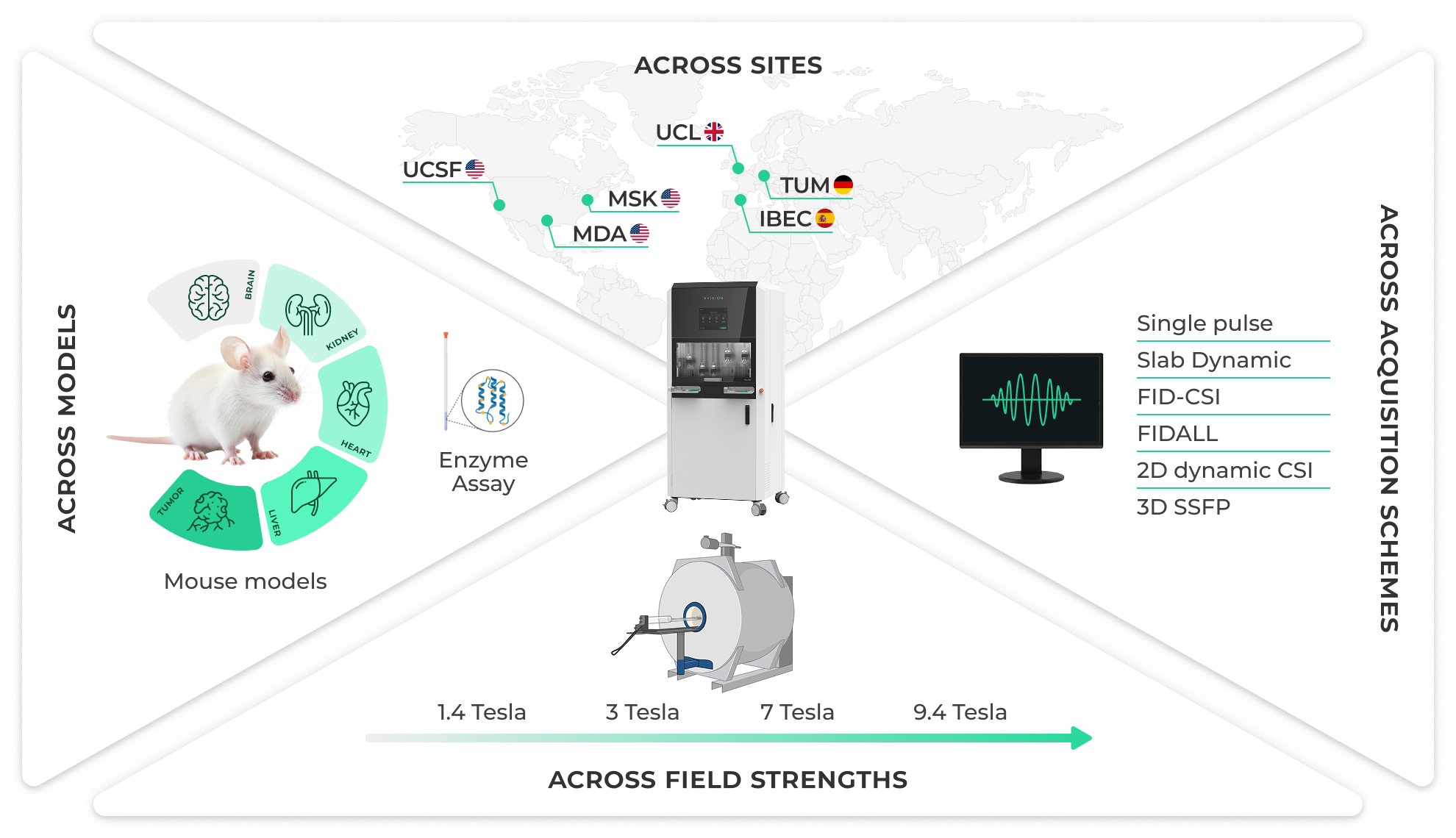
Hyperpolarized MRI can visualize metabolic processes that were previously inaccessible with other imaging techniques, yet producing the necessary imaging agents (also known as hyperpolarized agents) has long been slow, complex, and difficult to reproduce across multiple sites. To tackle these challenges, researchers at six leading centers in Europe and the U.S. partnered with NVision to use the POLARIS Preclinical polarizer and gain real-world experience with its workflow, exploring its potential to make hyperpolarized agent production faster, simpler, and more reliable. Across all sites, the system demonstrated not only the feasibility of standardized, high-throughput metabolic imaging, but also how a reproducible workflow, independent of field strength, imaging platform, or location, makes collaborative research possible. Workflows of this type could help support the path toward clinical translation, and pave the way for wider adoption of hyperpolarized metabolic imaging in both research and clinical settings.
Multi-site experience Across Six Centers
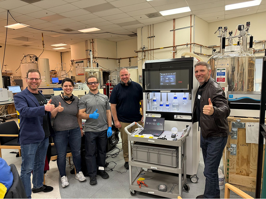
Two POLARIS systems were used across:
- Technical University of Munich (TUM), Germany
- Institute for Bioengineering of Catalonia (IBEC), Spain
- University of California San Francisco (UCSF), USA
- MD Anderson Cancer Center (MDA), USA
- University College London (UCL), UK
- Memorial Sloan Kettering Cancer Center (MSK), USA
Across the studies, 340 hyperpolarized doses were produced, of which 190 were used in vivo across 53 mice, with additional doses supporting dose characterization or in vitro studies. These in vitro experiments included time-resolved spectroscopy at 1.4 T to monitor pyruvate-to-lactate conversion under varying LDH enzyme activity levels. While in vivo scans were performed at multiple field strengths (3, 7, and 9.4 T). The analysis of the data was also standardized; for example, all in vivo data were analysed using NVision’s post processing tool, which enables peak fitting and metabolite mapping among other functions.
The average polarization level (~22%) and long T₁ values (~150 s) demonstrated a robust performance across multiple sites, field strengths and imaging platforms.
Site Highlights
Technical University of Munich (TUM), Germany
The Schilling Lab used POLARIS-generated doses to spatially map metabolism in the mouse heart and kidneys. This study highlighted the feasibility of high-resolution metabolic imaging across multiple organ systems with these doses.
Institute for Bioengineering of Catalonia (IBEC), Spain
In collaboration with Dr. Irene Marco Rius and her team, POLARIS Preclinical was integrated into studies of pyruvate-to-lactate conversion in the liver. Time-resolved spectra captured lactate production, demonstrating the system’s applicability for non-invasive dynamic metabolic assessments in hepatology.
University of California San Francisco (UCSF), USA
At UCSF — the first North American installation — doses were used in vivo to investigate healthy brain metabolism. Prof. Dan Vigneron summarized their joint experience, noting:
“In our recent experience, the new POLARIS Preclinical polarizer demonstrated excellent performance, reliability and safety through both quantitative testing and preclinical mouse studies. The ability to routinely generate sequentially many hyperpolarized doses through dozens of in vivo experiments and its ease of use indicates a very strong and important potential to advance the field of metabolic imaging in both preclinical and ultimately patient studies of human diseases addressing unmet clinical needs.”
MD Anderson Cancer Center (MDA), USA
At MD Anderson, POLARIS Preclinical enabled Prof James Bankson and his team to monitor metabolism in preclinical tumor models, providing a foundation for studying response-guided therapeutic strategies.
University College London (UCL), UK — CHARM Initiative
As part of the UK-Germany CHARM collaboration (Calibration of Hyperpolarized metAbolic MR), POLARIS Preclinical supported the Punwani lab and Prof Xavier Golay’s team at Gold Standard Phantoms in the development of calibration methods for future clinical translation. Over only three weeks, the system supported more than 100 hyperpolarization experiments, demonstrating strong robustness for high-volume standardization work.
Memorial Sloan Kettering Cancer Center (MSK), USA
At MSK, POLARIS Preclinical was fully deployed and is being used for preclinical cancer metabolism research to support the exploration of new therapeutic strategies. Read the full press release here.
A Step Forward for Accessible Hyperpolarization
This multi-site experience demonstrates that PHIP-based hyperpolarization, in the form of POLARIS Preclinical, can be reliably deployed across diverse institutions and MRI configurations. Nvision’s Head of Research, HP-MRI Applied Science, Myriam Chaumeil, reflected on what this means for the field:
“By dramatically simplifying the hyperpolarization workflow and improving user’s experience, POLARIS Preclinical enables research teams to generate hyperpolarized metabolic imaging data effortlessly. This strengthens feasibility and reproducibility, which in turn makes multicenter studies possible in short time frames, helping support the path toward much needed clinical translation.”
NVision is grateful for the collaboration with all of these research centers, and we look forward to continuing this shared effort to expand what metabolic imaging can achieve. Together.